CBCT: A Revolution in Oral and Maxillofacial Surgery Imaging
Oral and maxillofacial surgery has experienced significant advancements in recent years, including the introduction of Cone Beam Computed Tomography (CBCT). This cutting-edge imaging technology has revolutionized surgical planning and diagnostic accuracy for various procedures, from dental implants to orthognathic surgery. In this post, we will briefly introduce the topic of using CBCT in oral and maxillofacial surgery, explain what CBCT is, and how it differs from traditional 2D imaging methods. Additionally, we will provide an overview of the keywords discussed in the post.
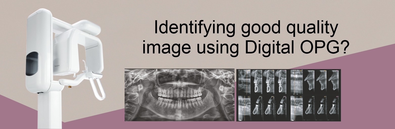
What is CBCT, and How Does It Differ from Traditional 2D Imaging Methods?
Cone Beam Computed Tomography (CBCT) is a three-dimensional (3D) imaging technology that uses a cone-shaped X-ray beam and a digital sensor to capture multiple images at various angles around the patient. These images are then reconstructed into a single 3D dataset, offering a comprehensive view of the patient’s anatomy.
In contrast, traditional 2D imaging methods, such as panoramic and periapical radiographs, only provide a limited perspective of the patient’s anatomy. While these methods have been widely used in dentistry and oral surgery, they often need more precision and depth to assess complex cases accurately.
The use of CBCT in oral and maxillofacial surgery offers several advantages over traditional 2D imaging, including:
- Enhanced diagnostic accuracy
- Improved surgical planning
- Minimized risk of complications
- Better patient outcomes
By delving into these topics, we will explore the numerous benefits and applications of CBCT in oral and maxillofacial surgery. Stay tuned for an in-depth discussion of how this innovative imaging technology transforms the field and improves patient outcomes.
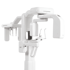
CBCT Technology
Cone Beam Computed Tomography (CBCT) is a cutting-edge imaging technology that uses a cone-shaped X-ray beam and a digital sensor to capture multiple images at various angles around the patient. The captured images are then reconstructed using advanced algorithms into a single 3D dataset, offering a comprehensive and detailed view of the patient’s dental anatomy, including bone structure, teeth, and surrounding soft tissues.
Benefits of Using CBCT in Oral and Maxillofacial Surgery
- Three-dimensional images: Unlike traditional 2D imaging methods, CBCT provides 3D images that enable oral and maxillofacial surgeons to visualize better and understand the complexities of the patient’s anatomy. This allows for a more accurate diagnosis and effective treatment planning.
- Enhanced diagnostic accuracy: CBCT technology provides high-resolution images with minimal distortion, resulting in a more accurate diagnosis of various conditions, such as impacted teeth, jaw discrepancies, and pathological lesions.
- Improved surgical planning: With the detailed information provided by CBCT, surgeons can develop precise and customized surgical plans, reducing the risk of complications and optimizing patient outcomes.
- Minimized radiation exposure: CBCT exposes patients to significantly less radiation than traditional CT scans while providing high-quality images.
CBCT vs. Traditional 2D Imaging Methods
Traditional 2D imaging methods, such as panoramic and periapical radiographs, have been widely used in dentistry and oral surgery for many years. However, these methods often need more precision and depth to assess complex cases accurately. CBCT overcomes these limitations by providing the following advantages:
- Comprehensive view: CBCT offers a complete 3D representation of the patient’s anatomy, allowing for a more thorough assessment of the structures involved.
- Greater detail: CBCT images provide more detailed information about bone quality, nerve location, and proximity of impacted teeth to adjacent structures, enabling surgeons to make better-informed decisions about surgical approaches and techniques.
- Reduced image distortion: CBCT technology minimizes distortion caused by overlapping structures, making it easier for surgeons to assess the patient’s anatomy accurately.
Dental Procedures that Benefit from CBCT
Cone Beam Computed Tomography (CBCT) has emerged as a revolutionary imaging technology in oral and maxillofacial surgery. By providing detailed three-dimensional (3D) images, CBCT allows surgeons better to understand the complexities of a patient’s anatomy and develop more precise treatment plans. This blog will discuss the different types of oral and maxillofacial surgical procedures that can benefit from CBCT, including dental implant surgery, orthognathic surgery, maxillofacial trauma, temporomandibular joint (TMJ) disorders, and oral pathology. Additionally, we will explain how CBCT can help in each of these procedures.
Dental Implant Surgery Dental implant surgery requires precise planning and execution to achieve successful outcomes. CBCT plays a critical role in this process by providing detailed information about bone quality, quantity, and the location of vital structures such as nerves and blood vessels. This information lets surgeons determine the optimal implant position, angulation, and depth, ultimately leading to more predictable and successful outcomes.
Orthognathic Surgery
Orthognathic surgery involves the correction of jaw discrepancies to improve function and facial aesthetics. CBCT is invaluable in diagnosing and planning these complex surgeries, as it enables surgeons to visualize skeletal structures and prepare the necessary bone movements with greater accuracy. Additionally, CBCT can be combined with digital simulation software, allowing surgeons to predict surgical outcomes and share these visualizations with patients.
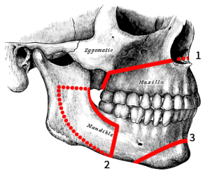
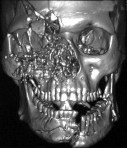
Maxillofacial Trauma
Maxillofacial trauma can result in fractures and dislocations of facial bones. Accurate diagnosis and surgical planning are crucial for successful treatment. CBCT provides high-resolution 3D images that help surgeons assess the extent and complexity of the injury, plan surgical approaches, and visualize the precise alignment of fractured segments during the surgery, leading to improved patient outcomes.
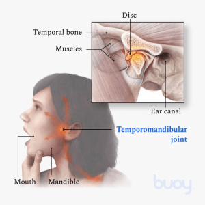
Temporomandibular Joint (TMJ) Disorders
CBCT imaging has proven beneficial in the assessment and management of TMJ disorders. It allows for a detailed evaluation of the bony structures of the joint, detecting degenerative changes and identifying the position and morphology of the articular disc. This information enables surgeons and other healthcare professionals to develop customized treatment plans and monitor therapy progress.
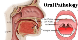
Oral Pathology
CBCT is a valuable tool for diagnosing and managing various pathologies affecting the jaws, such as cysts, tumors, and infections. The 3D images provide a detailed view of the lesion’s size, shape, extent, and relationship to adjacent structures. This information is crucial for determining the appropriate treatment approach and monitoring the healing process.
Advantages of CBCT in Oral & Maxillofacial Surgery
Greater Accuracy and Precision in Diagnosis and Treatment Planning
One of the critical advantages of CBCT is its ability to provide high-resolution three-dimensional (3D) images of the patient’s dental and maxillofacial anatomy. This comprehensive view enables surgeons to:
Assess complex cases more accurately: CBCT images offer a clear and detailed view of the patient’s anatomy, helping surgeons identify impacted teeth, jaw discrepancies, and pathological lesions with greater precision.
Develop customized treatment plans: The detailed information provided by CBCT allows surgeons to develop precise and individualized surgical plans, minimizing the risk of complications and optimizing patient outcomes.
Reduced Radiation Exposure Compared to Traditional CT scans
CBCT technology exposes patients to less radiation than traditional CT scans while providing high-quality images. This reduced radiation exposure is particularly beneficial for younger patients, pregnant women, and patients requiring multiple scans. As a result, CBCT is considered a safer imaging modality for a wide range of oral and maxillofacial procedures.
Faster and More Efficient Diagnosis and Treatment
CBCT imaging offers several advantages in terms of speed and efficiency:
- Faster image acquisition: CBCT scans can be completed in seconds, making them more convenient for patients and reducing the risk of image distortion caused by patient movement.
- Streamlined workflow: CBCT images can be easily integrated into digital workflows, allowing for efficient communication and collaboration between surgeons, dental laboratories, and other healthcare professionals.
- Immediate diagnosis and treatment planning: CBCT images can be assessed immediately after acquisition, enabling surgeons to diagnose conditions and develop treatment plans more quickly, ultimately leading to faster treatment and better patient outcomes.
Conclusion
In this post, we have explored the numerous advantages and applications of Cone Beam Computed Tomography (CBCT) in oral and maxillofacial surgery. To recap, CBCT offers:
- Greater accuracy and precision in diagnosis and treatment planning
- Reduced radiation exposure compared to traditional CT scans
- Faster and more efficient diagnosis and treatment
Genoray Papaya 3D Plus CBCT Machine has proven to be a game-changer in oral and maxillofacial surgery, enabling surgeons to provide better patient care and outcomes through accurate diagnosis and precise treatment planning. The integration of this cutting-edge imaging technology has set a new standard of care, transforming the way we approach complex surgical procedures.
Suppose you are a patient considering oral and maxillofacial surgery or a dental professional interested in learning more about the benefits of CBCT. In that case, I encourage you to speak with your oral and maxillofacial surgeon. They can provide valuable insight into how CBCT can be used to optimize your dental procedures and enhance the overall quality of care.
As an oral and maxillofacial surgeon, I am excited to see how CBCT technology will continue to shape the future of our field, and I look forward to sharing my experiences and insights with you.


Leave a comment