
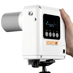
DC vs AC Dental X Rays
Among the numerous innovations, dental X-rays have undeniably revolutionized the landscape of dental diagnosis, enabling practitioners to delve beneath the surface for a more accurate, in-depth understanding of oral health conditions. The radiant power of dental imaging technology, combined with the science of radiography, has been instrumental in elevating the image quality and, subsequently, the precision of diagnoses.
Dental X-Rays: An Indispensable Tool for Dental Diagnosis:
Dental X-rays are more than just diagnostic tools; they are the bedrock of preventive dentistry. They enable dentists to identify potential oral health issues that may not be visible to the naked eye. Remember, they play a significant role in the planning and execution of complex dental procedures such as root canals and dental implants.
Radiography & Dental Imaging Technology: A Match Made in Diagnostic Heaven:
Unlike traditional film-based X-rays, digital X-rays are faster, more efficient, and offer superior image quality. These high-definition images can be enhanced, magnified, and even color-adjusted to understand the oral structures and underlying conditions better.
Striking the Balance: Image Quality & Radiation Exposure:
While dental X-rays are irreplaceable in dentistry, managing radiation exposure without compromising image quality is paramount. Advances in dental imaging technology have significantly reduced radiation exposure. Technologies like Direct Current (DC) dental X-rays generate a steady flow of electric current, which results in sharper images with less radiation exposure compared to their Alternating Current (AC) counterparts.
Understanding Dental X-rays
Dental X-rays, or radiographs, are the unsung heroes of modern dentistry. They enhance the accuracy of diagnoses and serve as an indispensable tool in the broader scope of treatment planning. As we delve deeper into dental imaging technology, let’s take a moment to comprehend the concept, purpose, and role of dental X-rays and the advancements that have shaped this vital technology.
Dental X-Rays: The See-Through Vision:
Dental X-rays are a type of picture that allows dentists to view the interior structures of your mouth, including areas that are not visible during a regular dental examination. The purpose of these radiographs is manifold. They help detect cavities hidden between teeth, reveal dental infections, showcase bone loss, and even help monitor a child’s dental development. By offering this inside view, dental X-rays act as a preventive mechanism, enabling early intervention and reducing the need for complex procedures in the future.
Role of Dental X-Rays in Dental Diagnosis & Treatment Planning:
The contribution of dental x-rays to dental diagnosis and treatment planning is monumental. Whether it’s assessing the health of the roots, checking the status of a developing tooth, or mapping the anatomy of your mouth before a surgical procedure, dental x-rays offer invaluable insights.
A Glimpse into the Advancements in Dental Imaging Technology:
From traditional film x-rays to digital x-rays and 3D imaging, dental radiography has come a long way. Digital radiography, in particular, offers benefits such as immediate image viewing, easy image enhancement for improved diagnosis, and significantly lower radiation exposure compared to traditional film x-rays.
DC (Direct Current) Dental X-rays
In the diverse realm of dental radiography, DC (Direct Current) dental x-rays have carved a niche for themselves, showcasing an impressive blend of image quality, operational efficiency, and patient safety.
- Unravelling DC Dental X-Rays:
DC dental x-rays refer to radiography where a Direct Current powers the X-ray machine. The primary distinguishing factor here is the constant and unidirectional flow of electric charge in DC dental x-rays, as opposed to the periodically reversing direction of electric current in Alternating Current (AC) x-rays. This consistent electrical output is crucial in determining the overall performance of the X-ray machine, affecting aspects like exposure time, image quality, and radiation dosage.
- Components and Functioning of DC X-Ray Machines:
DC dental X-ray machines have a generator, an X-ray tube, and a control panel. The generator produces the electrical power needed to create x-rays. The X-ray tube, which includes a cathode and an anode, is where the x-rays are produced. The control panel allows the operator to adjust various parameters, such as exposure time and voltage.
When the machine is powered on, the electrons from the cathode are accelerated toward the anode, producing x-rays. These x-rays then pass through the patient and create an image on the detector. The continuous, direct current ensures a constant rate of x-rays, resulting in more consistent and reliable imaging results.

Advantages and Disadvantages of DC Dental X-Rays:
- Image Quality Considerations:
One of the most significant advantages of DC dental x-rays is their superior image quality. Thanks to the constant current and voltage supplied by DC generators, there is less fluctuation in the X-ray output, reducing the potential for motion blur and enhancing image clarity.
- Radiation Exposure Considerations:
Regarding radiation exposure, DC dental x-rays typically deliver a lower dose than their AC counterparts, making them a more patient-friendly choice.

- AC (Alternating Current) Dental X-rays
These radiographic tools offer an interesting contrast to their DC counterparts and have played an integral role in shaping the modern dental diagnostic landscape. Let’s delve deeper into the concept, operation, and pros and cons of AC dental x-rays.
- Decoding AC Dental X-Rays:
AC dental x-rays operate on Alternating Current, characterized by an electric charge that periodically changes direction. This fundamental characteristic brings unique features to AC dental X-ray machines, affecting various parameters such as exposure time, image quality, and radiation dosage.
- Components and Functioning of AC X-Ray Machines:
Like DC X-ray machines, AC dental X-ray machines are composed of a generator, an X-ray tube, and a control panel. The generator creates the power needed for X-ray production. Inside the X-ray tube, x-rays are produced when high-speed electrons emitted from the cathode collide with the anode. The control panel allows for adjustments of different parameters, such as voltage and exposure time.
In an AC system, as the current changes direction, there is a slight pause between each cycle, causing a small fluctuation in the X-ray output. This can impact the overall image quality and exposure time.
Advantages and Disadvantages of AC Dental X-Rays:
- Image Quality Considerations:
While AC dental x-rays can produce high-quality images, the alternating nature of the current may cause slight inconsistencies in the X-ray output, potentially leading to minor motion blur in the picture. Therefore, utmost care is needed in operating AC X-ray machines to ensure optimum image quality.
- Radiation Exposure Considerations:
AC dental x-rays can emit a slightly higher radiation dose than DC x-rays due to the periodic reversal of the electric charge. However, this difference is minimal and, with appropriate safety measures, falls well within safe radiation exposure limits. The principle of ALARA (As Low As Reasonably Achievable) continues to guide dental professionals in optimizing the balance between diagnostic accuracy and patient safety.

Comparative Analysis: DC vs AC Dental X-rays
When it comes to dental imaging technology, it is crucial to understand the subtle yet significant differences between DC (Direct Current) and AC (Alternating Current) dental x-rays. While both are indispensable tools in the arsenal of modern dentistry, a comparative analysis provides valuable insights into their characteristics and their collective impact on dental diagnosis and treatment planning.

- Image Quality Comparison:
Regarding image quality, DC dental x-rays generally hold an edge over AC dental x-rays. The consistent and unidirectional flow of electric charge in DC dental x-rays results in a stable X-ray output, reducing the risk of image blurring and providing more precise and sharper images. On the other hand, the periodically reversing direction of electric current in AC dental x-rays can lead to minor inconsistencies in the X-ray output, resulting in slight motion blur in the images. However, AC dental x-rays can still deliver high-quality photos with careful operation and adjustment.
- Radiation Exposure Comparison:
Safety is paramount in any medical field, and dentistry is no exception. DC dental x-rays typically emit a lower radiation dose than AC dental x-rays. This is due to the steady current and voltage supplied by DC generators, which result in a more regulated X-ray beam and, thus, lower radiation exposure. However, it’s worth noting that the radiation dose from AC dental x-rays, while slightly higher, is still within safe limits, especially with appropriate safety measures.
- Impact on Dental Diagnosis and Treatment Planning:
DC and AC dental x-rays are pivotal in dental diagnosis and treatment planning. The high-resolution images obtained from both types of x-rays provide crucial insights into the internal structures of the mouth, thereby aiding in accurate diagnosis and effective treatment planning. The choice between DC and AC typically comes down to the specifics of the dental procedure, the needs of the patient, and the dentist’s preference.
In conclusion, both DC and AC dental x-rays bring their unique strengths. While DC x-rays offer superior image quality and lower radiation exposure, AC x-rays have been a longstanding, reliable tool in dental radiography. As dental professionals, the role is to continuously refine the understanding of these technologies and use them to enhance the standard of care for patients.
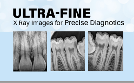
Ensuring Image Quality in Dental X-rays
Image quality is the lynchpin of dental radiography, significantly influencing the precision of diagnosis and treatment.
- Factors Influencing Image Quality in Dental X-Rays:
X-Ray Generator Parameters: The voltage and current settings on the X-ray generator directly impact the energy and quantity of the x-rays produced. The higher voltage generates more energy, leading to more penetration and less contrast, while the higher current has more x-rays, leading to a brighter image.
- Patient Positioning and Image Capture Technique:
Correct patient positioning and precise image capture technique are crucial in acquiring accurate and distortion-free images.
Image Processing: With digital x-rays, image processing plays a pivotal role in the final image quality. Adjustments like contrast enhancement, brightness control, and noise reduction can significantly improve the image’s diagnostic value.
- Quality Control Measures for Dental X-Ray Machines:
Maintaining high image quality standards requires a concerted effort and regular checks on the equipment and techniques. Here are some essential quality control measures:
Regular Calibration: Regular calibration of the X-ray generator ensures that it functions within its specified parameters.
Image Receptor Care: Digital sensors or X-ray film must be handled and stored to avoid any physical damage or exposure to unnecessary radiation.
Equipment Maintenance: Regular maintenance checks and immediate rectification of any identified issues help maintain the equipment’s optimal functioning and extend its lifespan.
Continuous Education: Regular training and updates on image capture techniques and image processing help the dental team stay abreast of best practices in dental radiography.
Radiation Safety Measures: Regularly checking and maintaining protective equipment, such as lead aprons, and ensuring the ALARA principle is being followed is crucial.
Managing Radiation Exposure in Dental X-rays
Radiation safety is of paramount importance in the field of dentistry. While dental x-rays significantly contribute to successful diagnosis and treatment planning, they also entail a degree of radiation exposure. Therefore, managing this exposure efficiently and effectively is critical to ensuring the safety of both patients and dental professionals.
- Importance of Radiation Safety in Dental Practice:
While the level of radiation exposure from dental x-rays is relatively low, no radiation dose can be considered entirely risk-free. Therefore, the onus is on dental professionals to ensure the safety of patients and staff through rigorous radiation safety measures. Adherence to these measures enhances patient safety and boosts the confidence and trust patients place in their dental care providers.
- Strategies for Minimizing Patient Radiation Exposure:
Minimizing radiation exposure is guided by the ALARA (As Low As Reasonably Achievable) principle. Here are some strategies to ensure patient safety:
Justification: Perform x-rays only when clinically necessary and with the patient’s informed consent.
Optimization: Use the lowest possible radiation dose that yields a diagnostically valuable image.
Digital X-rays: Digital x-rays expose patients to less radiation than traditional film x-rays.
Lead Aprons and Thyroid Collars: Protective equipment protects the patient’s body from unnecessary radiation.
Image Re-takes: Minimize image re-takes by proper patient positioning and accurate image capturing.
- Protective Measures for Dental Professionals:
In addition to protecting patients, dental professionals must also safeguard themselves against radiation exposure. Essential protective measures include:
Distance and Shielding: Maintain a safe distance from the X-ray beam and use protective barriers.
Personal Dosimeters: These devices measure the amount of radiation exposure and ensure it remains within safe limits.
Regular Training: Regular radiation safety training is crucial to stay updated on safety practices and regulations.
Design of the X-Ray Room: The design of the X-ray room should be such that it ensures maximum safety from scattered radiation.
- Dental Diagnosis and Treatment Planning with X-rays
Dental x-rays serve as our vision into the areas of the oral cavity hidden from the naked eye, aiding in diagnosing conditions and planning treatments with higher accuracy. Let’s examine the critical role of dental x-rays in this crucial process.
- Role of Dental X-Rays in Diagnosing Dental Conditions:
Dental x-rays are invaluable diagnostic tools that allow us to spot dental conditions at their earliest stages, even before they manifest visible symptoms. They help identify tooth decay, bone loss, tumors, and other abnormalities not visible during a routine dental examination. They also allow us to view the position and growth of teeth, which is particularly important in tracking the development of children’s teeth and planning orthodontic treatments.
- Application of Dental X-Rays in Treatment Planning:
Once a dental condition has been accurately diagnosed, dental x-rays are essential in treatment planning. Providing a detailed image of the patient’s teeth and surrounding structures enables us to devise a tailored treatment plan that addresses each patient’s individual needs. For instance, in restorative procedures like root canals or dental implants, x-rays guide us in determining the exact anatomy and choosing the best approach for treatment.
- Importance of Accurate and Reliable X-Ray Images:
Accuracy and reliability in X-ray images are vital. Inaccurate or unreliable photos can lead to misdiagnosis, ineffective treatment planning, and, ultimately, a compromise on the quality of care provided to the patient. As dental professionals, we commit to ensuring that the X-ray images we work with offer a proper, detailed, and clear view of the patient’s oral health condition.

Conclusion

Navigating through the world of dental x-rays can be a challenging task, filled with technical jargon and complex decision-making. Yet, with a deeper understanding of the concepts and considerations, one can appreciate the intricate dynamics and make informed decisions.
Recap of the Key Points Discussed:
Throughout our exploration, we have delved into the nuances of Dental X-Rays, touching upon their importance in dental diagnosis and treatment planning. We have explored the distinctions between DC and AC dental x-rays and discussed the impact of various factors on image quality and radiation exposure.
Considerations for Choosing between DC and AC Dental X-Rays:
Choosing between DC and AC dental x-rays is not a simple case of “one size fits all.” The choice depends on numerous factors, including clinical needs, patient characteristics, each system’s specific advantages and disadvantages, and the dentist’s comfort and familiarity with the equipment. The key to this choice is to balance obtaining high-quality images and minimizing radiation exposure.
Emphasis on Image Quality, Radiation Exposure, and Their Impact on Dental Diagnosis:
Finally, we cannot stress enough the significance of image quality and radiation safety in dental practice. They are not mere technical parameters but the very foundation of effective dental diagnosis and treatment planning. High-quality images allow for accurate diagnosis, while low radiation exposure ensures patient and practitioner safety. They are crucial components of the dental X-ray process and should be given the importance they deserve.


Leave a comment