The Emergence of Cone Beam Computed Tomography (CBCT) in Dentistry & Its Significance
Cone beam Computed Tomography (CBCT)
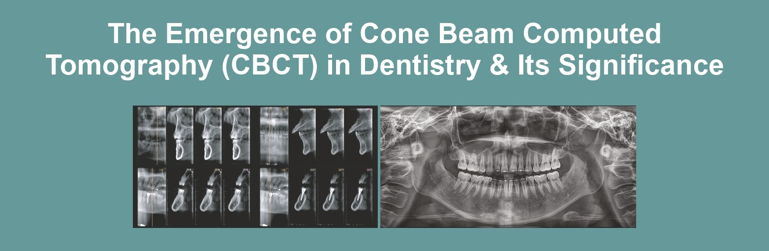
Computed tomography is an advanced imaging modality that has high clinical application in the field of dentistry. It is a radiographic imaging method that allows accurate, three-dimensional imaging of hard and soft tissue structures. A CBCT scanner uses a collimated x-ray source that produces a cone or pyramid-shaped beam of radiation, which produces a sequence of discrete images using a digital detector.

The First commercial CBCT unit marketed for dental use was introduced in Europe in 1999. Currently, the availability of this technology is increasing in many dental institutes and diagnostic centres across the country. CBCT is providing dental clinicians a modality for 3D representation of maxillofacial structures which in turn has reduced dependence on CT.
2D imaging to 3D imaging in Dental Practice
Imaging is an important aspect of the diagnosis and management of a patient. Although new imaging techniques are introduced to conventional 2D radiographs, panoramic or occlusal radiographs remain a commonly used modality for primary diagnosis. But 2D imaging has limitations like magnification, distortion, and superimposition. On the other side multiplanar imaging has provided the best diagnostic approach. The volume of data that is acquired during the CBCT scan is stored, reformatted, and realigned. The biggest advantage of CBCT imaging is the avoidance of superimposition.
With multiplanar imaging, images can be recreated in different planes. Images can be displayed in sagittal, axial, or coronal views. Also, this imaging can be used for specific areas like TMJ, which increases diagnostic efficiency in an unparalleled way.
What is CBCT used for in Dentistry?
The clinical application of CBCT imaging in the oral and maxillofacial area is vast. Multiplanar imaging in CBCT has provided a highly useful technique for the diagnosis of any bone pathology or any developmental anomaly in the Oro facial region.
CBCT in dentistry helps in the diagnosis of fractures, their size, extent, and location to assess their relation to vital structures. Also, CBCT is preferred for object localization, impacted teeth, supernumerary teeth, or any developmental anomalies.
Pre-surgical planning and post-operative evaluation can be easily done using CBCT
CBCT applications in Dentistry:
Is CBCT used for orthodontics?
CBCT is recommended in cases where diagnostic information provided by conventional radiography is not sufficient. Hence it’s a useful tool for patients with cleft palate, facial asymmetry, large open bites, and planning of orthognathic surgery. CBCT in orthodontics provides valuable information for the placement of endo osseous implants or temporary anchorage devices.
Use of CBCT for implants and endodontic treatment
CBCT use in dentistry plays an important role in the analysis of benign and malignant lesions and is the choice of technology for assessment of fractures of a tooth for identification and measurement of a periapical lesion to differentiate solid from fluid-filled lesions and very helpful even in the RCT.
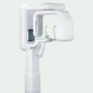
Genoray Papaya 3D plus:
 The CBCT machine used commonly in Dentistry is Genoray Papaya 3D Plus. This is the future of dental imaging which is a combination of 3D CT, Panoramic and Cephalometric to meet all diagnostic needs. The versatile imaging capability provides the user with accurate information for all dental treatments, especially implant placement. There are 3 different dedicated CMOS flat panel sensors for 3 different needs, so it gives long-lasting performance and long life as there is no overload on a single sensor for both 2D and 3Dimages
Features of Genoray Papaya 3D plus
It has a smart metal artifact removal feature for more clarity. Also, the innovative sensor technology used in this machine acquires images in just seconds, thus preventing patient movement and increasing image quality. The versatile imaging capability provided the user with accurate information giving a zero error image.
What are 3 limitations of use of CBCT imaging?
These include
- Higher doses than two-dimensional imaging;
- The inability to accurately represent the internal structure of soft tissues and soft-tissue lesions
- Limited contrast resolution compared to CT
Conclusion:
3D imaging has made complex craniofacial structures more accessible for examination and also helps in the early and accurate diagnosis of lesions. CBCT has reduced dependence on super specialty setup like CT fo3D imaging of oral and maxillofacial.

Leave a comment